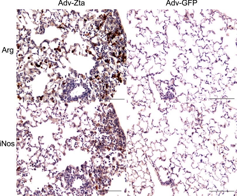
Fig. 5.
Alternative activation of macrophages by Zta. Macrophage activation status was determined by immunostaining histological sections of formalin-fixed, paraffin-embedded lung tissue for iNOS (M1) or arginase (M2). Brown staining marked cells positive for arginase (top) or iNOS (bottom). Adjacent lung sections from Adv-Zta (left)- and Adv-GFP (right)-treated mice killed on day 7 are shown. The scale bar indicates 100 μm.

