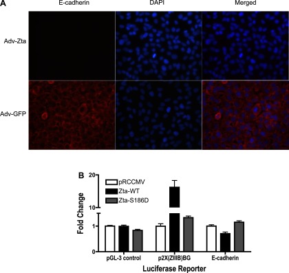
Fig. 9.
Repression of E-cadherin expression in lung epithelial cells by Zta. A: immunofluorescent detection of E-cadherin in A549 cells after infection with recombinant adenoviruses. A549 cells grown in chamber slides were infected with Adv-Zta (top) or Adv-GFP (bottom) at 20 moi. Three days postinfection, the cells were processed for immunofluorescent detection of E-cadherin (left). Cells were visualized with DAPI nuclear stain (middle). The merged images (right) demonstrate that Adv-Zta represses expression of E-cadherin in A549 cells (top), whereas Adv-GFP does not (bottom). B: repression of the E-cadherin promoter in lung epithelial cells by Zta. Mouse lung epithelial cells (C10) were cotransfected with firefly luciferase reporter constructs [pGL-3 control (negative control), p2X(ZIIIB)BG (positive control), and E-cadherin (pGL2Basic-EcadK1, test)], Zta-expressing plasmids [pRCCMV (empty vector), Zta-WT (wild-type Zta), and Zta-S186D (mutant Zta)], and pRL-TK-luciferase as an internal transfection efficiency control (see Fig. 7). The graph shows the mean fold change ± SE in firefly/renilla luciferase activity from 3 independent transfection assays performed in duplicate for the indicated firefly luciferase reporter construct. For each reporter plasmid, the mean firefly/renilla luciferase ratio in the presence of empty vector (pRCCMV) was normalized to 1. Induction of the 2X(ZIIIB)BG promoter and repression of the E-cadherin promoter by wild-type Zta is significant (P < 0.0001 and P = 0.0041).

