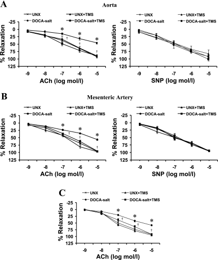
Fig. 4.
TMS prevents DOCA-salt-induced endothelial dysfunction in rats. Endothelial function was examined by measuring the response of the aorta (A) and mesenteric arteries (B) to increasing concentrations of ACh (10−9–10−5 mol/l) following constriction with PE (10−5 mol/l). Endothelium-independent vasodilation was examined by measuring the relaxation response to sodium nitroprusside (SNP; 10−9–10−5 mol/l) after the vessels were constricted with PE (10−5 mol/l). C: to determine the contribution of endothelium-derived hyperpolarizing factor (EDHF)-mediated relaxation, the response to ACh (10−9–10−5 mol/l) was determined in the presence of indomethacin (10−5 mol/l) and NG-nitro-l-arginine methyl ester (l-NAME; 10−4 mol/l). A combination of indomethacin and l-NAME was added to the myograph chamber ∼10 min before constriction with PE. Relaxation is expressed as a percentage of the contraction by PE. Data are means ± SE (n = 5). *P < 0.05, UNX vs. DOCA-salt. †P < 0.05, DOCA-salt vs. DOCA-salt + TMS.

