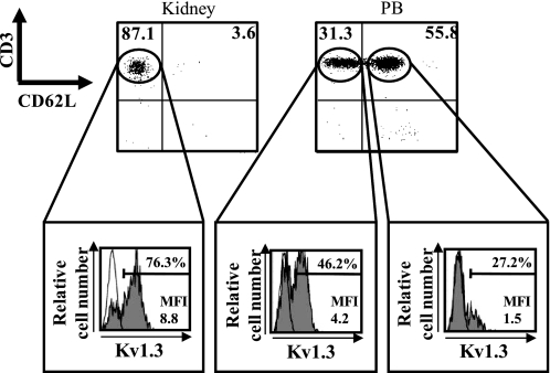
Fig. 5.
Expression of the Kv1.3 channel on CD62L− T cells (corresponding to effector memory T cells) and CD62L+ T cells (corresponding to naïve T cells and central memory T cells). αβ/γδTCR+ T cells isolated from kidney and peripheral blood (PB) in the vehicle group on day 7 were stained with rabbit anti-Kv1.3 polyclonal Ab conjugated with FITC, CD3 mAb conjugated with PE-Cy5, and CD62L mAb conjugated with phycoerythin (PE). Expression of Kv1.3 was analyzed after gating for CD62L− T cell and CD62L+ T-cell subpopulations. Numbers in the top left and top right quadrants are percentages of CD62L− T cells and of CD62L+ T cells, respectively. Mean fluorescence intensity (MFI) levels are shown for each histogram. Shaded histograms show the level of expression of Kv1.3 measured with FITC-conjugated polyclonal rabbit anti-Kv1.3 antibody. Open histograms represent staining obtained with the FITC-conjugated rabbit IgG isotype control.

