Fig. 3.
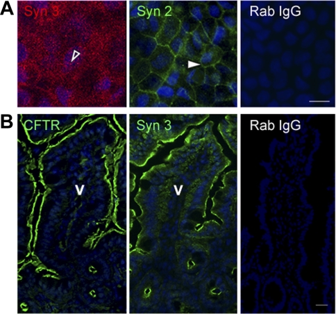
Polarized distribution of syntaxin 2 and 3 in the intestine. Confluent polarized Caco-2BBe cells (A) and cryostat sections of mouse jejunum (B) were fixed and immunofluorescence performed as described in materials and methods. A: en face image taken at the level of the brush border shows the apical distribution of syntaxin 3 (open arrowhead). Image taken at basal level shows distribution of syntaxin 2 (Syn 2, solid arrowhead). B: staining for CFTR (green) and syntaxin 3 (green) in the apical domain of crypt and villus sections of mouse jejunum. Control labeling of cells (A) and tissue sections (B) with rabbit IgG (Rab IgG) antibody is shown. Hoechst nuclear stain labels the nuclei (blue) V, villus. Scale bar, 10 μm.

