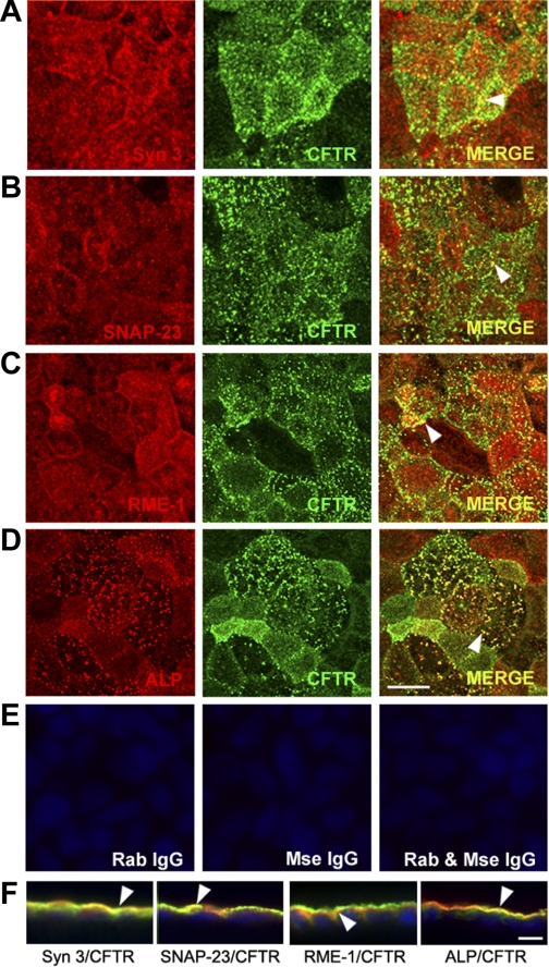
Fig. 4.
Subcellular distribution of endogenous CFTR and apical recycling and exocytic proteins in polarized Caco-2BBe cells. Confluent polarized Caco-2BBe cells were fixed, double-label immunofluorescence was performed, and cells were examined by confocal microscopy. A–D: en face images taken just above the level of the brush border show the distribution of syntaxin 3 (red, A), SNAP-23 (red, B), rme-1 (red, C), alkaline phosphatase (ALP, red, D), and CFTR (green, A–D). Merged images show colocalization (merge, yellow, arrowhead). E: control labeling with rabbit (Rab) and/or mouse (Mse) IgG antibodies. F: merged images of xz vertical sections of immunolabeled cells. Arrowheads indicate colocalization. Hoechst nuclear stain labels nuclei blue. Scale bar, 10 μm.

