Fig. 5.
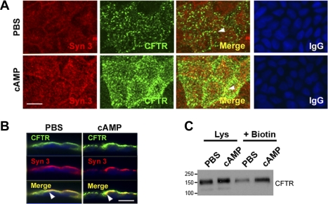
cAMP stimulates apical recruitment of syntaxin 3 and CFTR in Caco-2BBe cells. Confluent monolayers of Caco-2BBe cells were treated with PBS or 1 mM cAMP. Cells were fixed, immunolabeled, and examined by confocal microscopy. A: en face views show the distribution of syntaxin 3 (red) and CFTR (green), and merged images show areas of colocalization (yellow) in PBS (top)- or cAMP (bottom)-treated cells. Control staining with relevant IgG antibodies is shown. B: images of vertical xz sections of immunolabeled cells show CFTR (green) and syntaxin 3 (red), and merged images (yellow) show colocalization (arrowheads). Scale bar, 10 μm. C: after PBS or cAMP treatment surface proteins were detected by sulfo-NHS-SS-biotin labeling and analyzed by Western blot as described in materials and methods. Cell lysates (Lys, 20 μg protein) and equivalent loads of surface biotinylated (+Biotin) proteins were resolved by SDS-PAGE and immunoblotted to detect CFTR. Molecular mass standards (kDa) are indicated.

