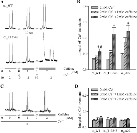
Fig. 6.
Ca2+ release induced by extracellular electrical stimuli in α1SWT- and α1ST1354S-expressing myotubes. A and C: Ca2+ transients from myotubes were elicited by 3 extracellular electrical pulses during sequential 30-s application of 0, 1, and 2 mM caffeine (indicated by gray bars of different width) in extracellular solutions containing 2 mM Ca2+ (A) and 0 mM Ca2+ (C, bottom lines). Between the pulse trains myotubes were incubated for 2 min with extracellular caffeine-free solution containing 10 mM Ca2+ for SR reloading (A). B: integral of Ca2+ transients in 2 mM extracellular Ca2+ as a measure for total Ca2+ release in response to one action potential. A trend towards higher Ca2+ release with constructs α1ST1354S (n = 23) and α1SΔ29 (n = 29) compared with α1SWT (n = 19) is observable under 1 mM caffeine and is significant under 2 mM caffeine (*P, #P < 0.05, respectively). D: integral of Ca2+ transients in 0 mM extracellular Ca2+ shows no difference between α1SWT (n = 13), α1ST1354S (n = 6), and α1SΔ29 (n = 14) in relation to different caffeine exposures.

