
Figure 3.
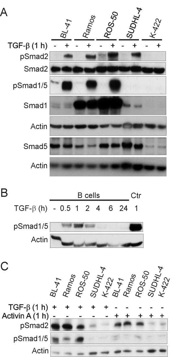
TGF-β sensitive cell lines signal through Smad1/5 in addition to Smad2. Cell lines were stimulated with or without TGF-β (A) and with activin A or TGF-β (C) for 1 h, and primary B cells were stimulated with or without TGF-β for 30 min, 1, 2, 4, 6 and 24 h (B), lysed, and subjected to western immunoblotting analysis, with the indicated primary antibodies. Staining with an anti-phospho-Smad1/5 antibody was applied to confirm that Smad8 was not involved (data not shown). The positive control (Ctr) in Fig. 3B is BL-41 total cell lysate. Presented is one representative blot out of three with one representative actin loading control.