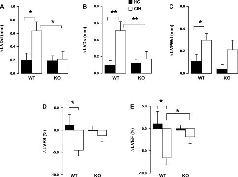
Fig. 2.
Changes (Δ) in echocardiographic indexes at week 8 over baseline. Values are means ± SE; N = 15 each group. A and B: changes of left ventricular (LV) diastolic (ΔLVDd; A) and systolic (ΔLVDs; B) dimension at week 8 over baseline. C: changes of LV posterior wall diastolic thickness (ΔLVPWd) at week 8 over baseline. D: changes of LV fractional shortening (ΔLVFS). E: changes of LV ejection fraction (ΔLVEF) at week 8 over baseline. *P < 0.05; **P < 0.01. Significance of interaction between genotype (WT or KO) and exposure (HC or CIH): for LVDd, P < 0.05; for LVDs, P = 0.0074; nonsignificant (NS) for LVPWd, LVFS, and LVEF.

