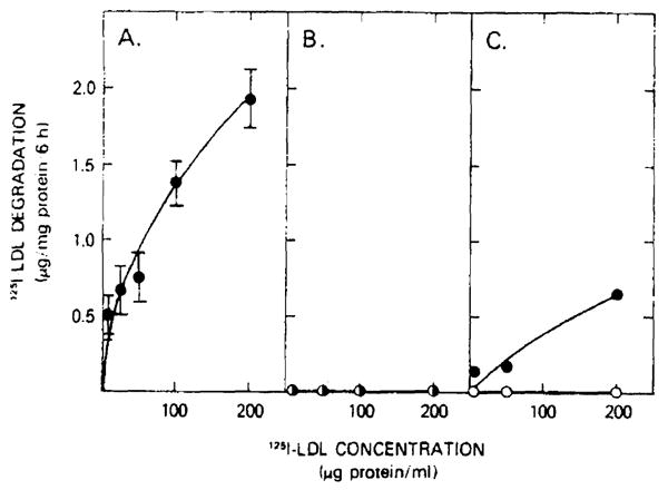
Fig. 5.

125I-labeled LDL degradation by hepatocytes from normal and homozygous familial hypercholesterolemia patients. The media from the normal hepatocytes (panel A), patient 1 hepatocytes (panel B), and from patient 2 (panel C) were harvested after a 6-h incubation with the indicated concentrations of 125I-labeled LDL. The non-chloroform-extractable 125I-label counts in the media (trichloroacetic acid supernatant) defined the degradation as outlined in Methods. Hepatocytes were incubated for 48 h prior to the exposure to 125I-labeled LDL in either the absence (○) or presence (●) of 2 mg unlabeled LDL protein/ml. Values for normal hepatocytes represent the mean ± S.E. for five different hepatocyte preparations.
