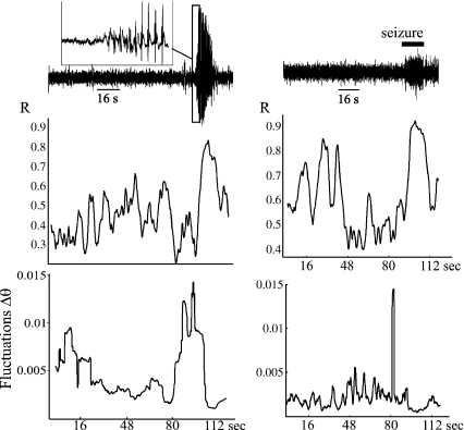
Fig. 2.
Growth of the fluctuations in phase difference between two MEG sensors in patient 4, with absence epilepsy (left), and patient 5, with frontal lobe epilepsy (right). The two sensors were located on the left frontal cortex (left) and in the central midline (right). Upper traces are the MEG recording in one sensor, and the inset denotes the start of the spike-and-wave absence seizure. Graphs show the phase synchrony index (R) and the fluctuations in phase difference (evaluated at 5 ± 2 Hz and 30 ± 2 Hz, respectively) showing a similar pattern to that revealed in Fig. 1. Note, however, that the absolute minimum of the R values coinciding with the increase in fluctuations is not clearly seen in the right-hand side case. Time scale (x-axes) is the same for allpanels in each column

