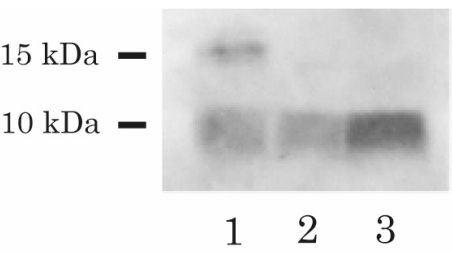
Figure 3.
Immunoblot of serum IGF-II. Lane 1 and Lane 2 show the pre- and postoperative blots for the patient; Lane 3 shows the blot for the normal subject. Western blot analysis was performed with the anti-IGF-II antibody (Upstate Biochemistry), and high molecular weight IGF-II (15 kDa) was detected in the preoperative serum but was not detected in the postoperative serum.

