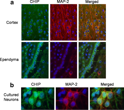
Fig. 1.
CHIP immunoreactivity in brain sections from adult male Long-Evans rats (a). Coronal sections were taken from the cortical and ependymal cell regions. The sections were stained with anti-CHIP antibody (green) and anti-MAP2 antibody (red) along with the nuclear counterstain bisbenzimide (blue). Merged images are shown in panels on the far right. CHIP immunocytochemical staining of cultured neurons shows reactivity in both neurons and astrocytes (b). Cultured neurons (10 days in vitro) were stained for CHIP (green) and MAP-2 (red) along with nuclear counterstain, bisbenzimide (blue). A merged image is shown in the panel on the far right

