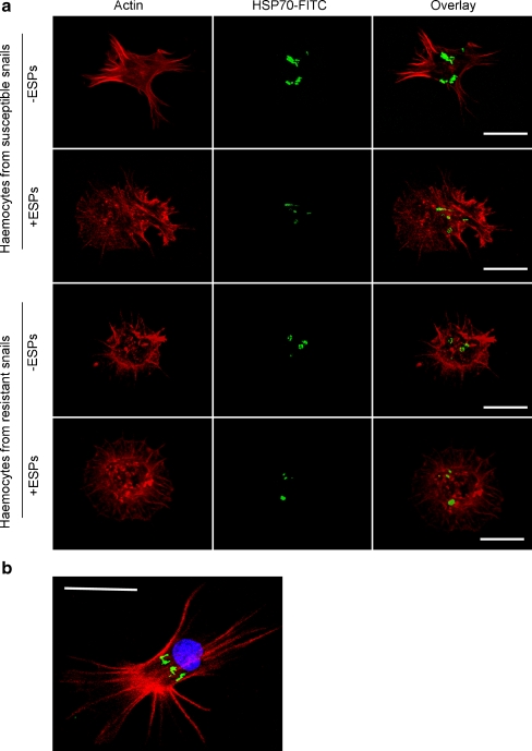
Fig. 5.
Cellular distribution and levels of HSP70 in B. glabrata haemocytes exposed or not exposed to S. mansoni ESPs. a Schistosome-susceptible and schistosome-resistant snail haemocytes were challenged with (+) and without (−) 20 μg/ml ESPs in CBSS for 1 h on glass coverslips before being fixed and subsequently stained with anti-HSP70 primary antibodies and FITC-conjugated secondary antibodies (represented by green); haemocytes were also incubated in rhodamine phalloidin (red) to visualise filamentous actin. Panel b shows a typical haemocyte not exposed to ESPs, additionally stained with DAPI (blue) to visualise the nucleus. Haemocytes were observed with a Leica laser scanning confocal microscope; images show z-axis projections in average pixel brightness mode that were captured using Leica software. The results shown are characteristic of those obtained in at least three independent experiments (bar 10 μm)

