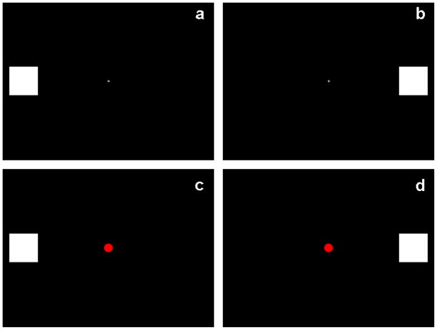
Figure 1.
Visual stimuli. The visual stimuli consisted of white squares (visual angle 2×2 degrees) presented on a black background for 82ms on the horizontal meridian, 6.79 degrees lateral to a central fixation cross. On target trials, the central fixation cross transformed into a red dot. 300 trials were presented, which delivered in 4 experimental blocks of 30 LVF non-target trials (panel a), 30 RVF non-target trials (panel b), 8 LVF target trials (panel c) and 7 RVF target trials (panel d), randomly presented. Participants were instructed to maintain their gaze on the central fixation cross and count the number of targets they saw.

