
Figure 3.
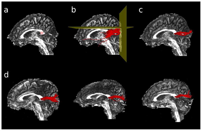
Extraction of the visual fibers of the corpus callosum with DTI tractography. A region-of-interest was first manually drawn in the splenium of the corpus callosum, on the midsagittal slice of each participants’ Fractional Anisotropy image (panel a). Voxels defined by the ROI were used as seedpoints for deterministic tractography. Fibers were excluded if they passed through the axial slice immediately dorsal to the corpus callosum, or if they failed to pass through the coronal slice defined by the posterior edge of the parieto-occipital fasciculus (panel b). The remaining fibers constituted the corpus fibers connecting the primary and secondary visual cortices bilaterally (panel c). Panel d shows the extracted visual corpus fibers for three representative individuals.