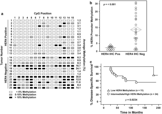
Figure 4.
Tumor hypermethylation of the HER4 promoter predicts breast cancer patient outcome. The patient population and HER4 expression analysis by immunohistochemistry (IHC) are described elsewhere (Thor et al., 2009). (a) Bisulfite-treated primary breast tumor DNA was analyzed by pyrosequencing as described in Figure 2d. DNA was isolated from 50 μm sections of formalin fixed and paraffin embedded (FFPE) primary breast tumors and bisulfite treated at EpigenDx. The position and extent of methylation for each site in 13 HER4-positive and 12 HER4-negative tumors is indicated. (b) Box plot of HER4 promoter methylation in HER4-positive (n=13) and HER4-negative (n=22) primary breast tumors (P<0.001). (c) Kaplan–Meier survival curves for disease-specific survival (DSS). HER4 methylation <3% (triangles; n=11) vs ⩾3% (circles; n=24) (P=0.0234) For Kaplan–Meier survival curves HER4 methylation determined by pyrosequencing was dichotomized at <3% vs ⩾3%. Outcomes included DSS defined as the number of months between the diagnosis date and the date of death from breast cancer.

