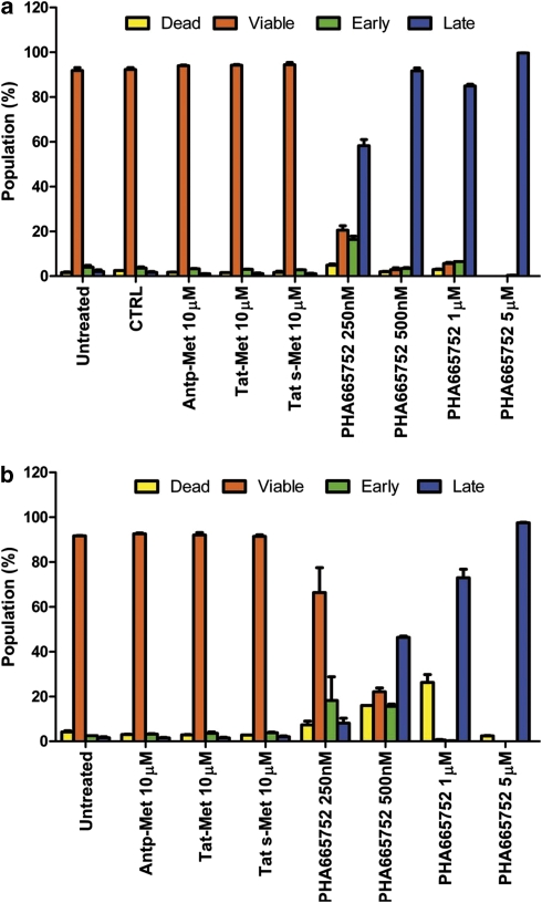
Figure 3.
Met docking site peptides do not induce toxicity or apoptosis in endothelial cells. In the apoptosis assay, HUVECs were plated onto six-well plates and were treated with either the Antp-Met peptide (10 μ), Tat-Met peptide (10 μ) or Tat s-Met peptide (10 μ) or with increasing concentrations of PHA-665752 (250 and 500 n, and 1 and 5 μ). Vehicle (dimethyl sulfoxide) and growth medium alone were used as controls, and HGF 50 ng/ml (panel a) or 10% FBS (panel b) were added to the growth medium. After 2 h, cells were recovered, washed and stained with two markers, Annexin V and 7-amino-actinomycin D, used to detect viable, dead or cells undergoing apoptosis. After only 2 h, 250 n PHA-665752 induced late apoptosis in about 60% of endothelial cells in HGF-containing media (a); on the contrary, 10 μ peptides showed an high percentage of viable endothelial cells (about 95% of the population; a). The toxicity of PHA-665752 was marked both in the presence of HGF (a) or FBS (b). The experiment was performed three times and each condition was in triplicate.

