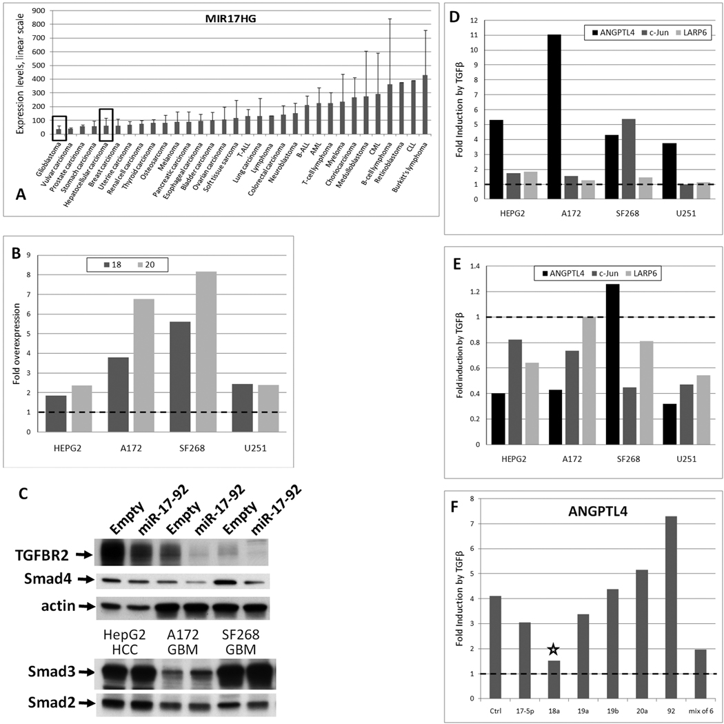
Figure 6. miR-17~92 impairs gene regulation by TGFβ.
A. Expression levels of MIR17HG across tumor types from the Wooster dataset. Error bars represent standard deviation. T-ALL denotes acute T-cell lymphoblastic leukemia, B-ALL - acute B-cell lymphoblastic leukemia, AML - acute myeloid leukemia, CML - chronic myeloid leukemia, CLL - chronic lymphoblastic leukemia. B. Fold overexpression of miR-18 and -20 in miR-17~92-tranduced glioblastoma (GBM) and hepatocellular carcinoma (HCC) cell lines relative to empty vector-transduced cells, as measured by qPCR. C. Expression levels of TGFBR2 and Smad proteins in cell lines depicted in the previous panel, as measured by Western blotting. D. Fold induction by TGFβ of three transcripts in the same cell lines as measured by qPCR. Treatment with TGFβ was carried out as in Figure 2. E. Comparison by qPCR of TGFβ effects on gene expression in miR-17~92-transduced cells relative to empty vector-transduced cells. F. Comparison by qPCR of TGFβ effects on ANGPTL4 in control (Ctrl) and miR-17~92 mimic-transfected A172 cells.

