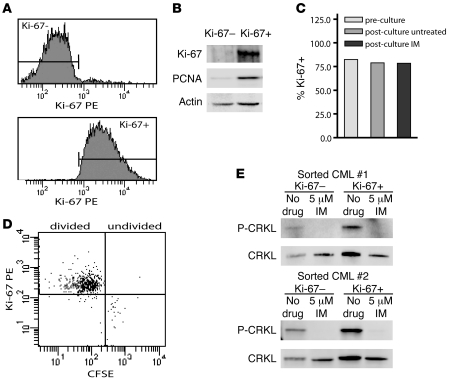
Figure 3. Inhibition of phospho-CRKL by imatinib in quiescent and cycling CML cells.
(A) Lin–Ki-67– and Lin–Ki-67+ CML cells were FACS sorted into quiescent and cycling fractions, respectively. Representative histograms are shown. (B) Immunoblots of Ki-67 and PCNA in sorted Lin–Ki-67– and Lin–Ki-67+ CML cells were used to confirm sort purity, as indicated by exclusive expression of these proteins in the cycling fraction. Representative blots are shown. Actin is included as a loading control. (C) Ki-67 expression was evaluated by FACS prior to and following 4-hour culture with or without 5 μM imatinib. (D) Lin– cells stained with CFSE and cultured for 72 hours with 5 μM imatinib were analyzed for Ki-67 expression. Ki-67 expression is shown in undivided (CFSEhi) cells and in cells having undergone 1 or more divisions (CFSElo/mod). (E) Phospho-CRKL immunoblots of quiescent versus cycling cells treated 4 hours with or without imatinib are shown for 2 newly diagnosed CML samples. Total CRKL is included as a loading control.

