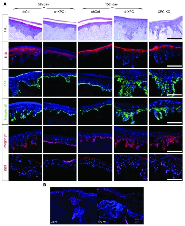
Figure 4. Knockdown of XPC results in epithelial hyperplasia.
Epidermis reconstructed with XPC-KC, shCtrl-, or shXPC-transduced keratinocytes on days 5 and 15 after transduction. (A) Architectures of epidermis were evaluated with H&E staining. Proliferation and differentiation status of epidermis were assessed using immunofluorescence staining of K10, K14, integrin α6, integrin β1, and Ki67. The nuclei were marked in blue with DAPI. (B) High proliferation detected with Ki67 staining in a large extension of rete pegs in epidermis reconstructed with XPCKD cells and XPC-KC. Scale bars: 200 μm.

