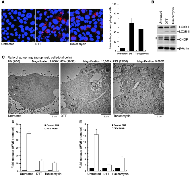
Figure 7. Suppression of the HCV- and DEV-PAMP RNA–induced IFNB promoter activation by UPR inducers.
(A–C) Huh7/RFP-LC3 cells were left untreated or treated with 2 mM DTT or 4 μg/ml tunicamycin for 6 hours, and then subjected to quantification of RFP-LC3–labeled puncta structure (scale bars: 10 μm) (A), Western blot analysis of the expressions of indicated proteins (B), or TEM analysis of autophagic vacuoles (C). The asterisk in B indicates the nonspecific background signal. An uncropped image of B is shown in Supplemental Figure 9, right panel. The ratio of autophagy in C was determined as described in Figure 6B. (D) Huh7/RFP-LC3 cells were transfected with the pIFN-β/Fluc promoter, followed by transfection with HCV PAMP RNA. The transfected cells were then treated with DTT or tunicamycin as described in A prior to assessment of IFNB promoter activation. The fold increase in IFNB promoter of the PAMP RNA–transfected cells was determined by normalization to the basal level of the control RNA–transfected cells. (E) The effects of DTT and tunicamycin on DEV PAMP RNA–mediated IFNB promoter reporter induction were determined as described in D. Data represent mean ± SEM (n = 3) (A, D, and E).

