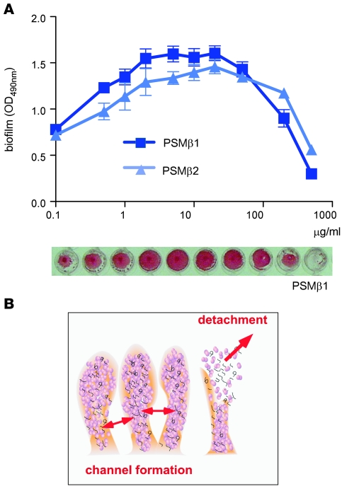
Figure 3. S. epidermidis in vitro biofilm formation under influence of PSMβ peptides.
(A) Biofilm formation in microtiter plates (24 hours, 37°C). PSMβ peptides at different concentrations were added at the time of inoculation with S. epidermidis agr (devoid of PSMs). Biofilms were made visible using safranin staining (see example for PSMβ1 at the bottom), and biofilm formation was measured using an ELISA reader. Error bars depict mean ± SEM. (B) Schematic presentation of biofilm cell-cell disruptive processes leading to channel formation and detachment.

