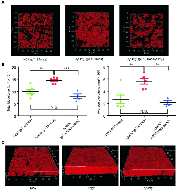
Figure 6. Role of PSMβ peptides and agr in S. epidermidis biofilm development: channel formation and biofilm expansion.
(A) CLSM pictures of S. epidermidis 1457 WT, isogenic psmβ mutant, and psmβ-complemented strains. The WT and psmβ mutant strains were transformed with the control plasmid pT181mcs to ensure comparability. Static biofilms were grown for 24 hours, stained with propidium iodide, and imaged using CLSM. View is from the top. (B) Analyses of total and average biovolumes, which are measures of total biofilm and degree of channel formation, respectively, using IMARIS software of the biofilm samples shown in part A. Note that increased average biofilm volume corresponds to decreased channel formation. **P < 0.01; ***P < 0.001, 1-way ANOVA with Bonferroni’s post tests. Error bars depict mean ± SEM. (C) CLSM pictures of S. epidermidis 1457 WT, isogenic agr, and psmβ operon deletion mutant biofilms. Growth conditions are as in A.

