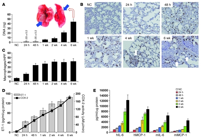
Figure 5. Tumor cells contribute to early macrophage infiltration and inflammation in the lung.
(A) Human 12p genomic DNA was detected by qRT-PCR DNA of 4 μg genomic DNA extracted from dissected lungs at the indicated time points after tail vein injection of 2 × 10⁶ UMUC3 cells/100 μl phenol red–free medium. Bars represent mean ± SEM of the amount of 12p DNA (ng) detected in lungs of 4 animals/group. Inset: Example of mouse lungs with visual metastases at 6 weeks after tail vein inoculation of UMUC3 cells. (B) Representative immunostaining (IHC) with macrophage marker mac2 antibody to assess number of macrophages infiltrating the lungs from normal lungs and lungs from animals injected with UMUC3, at the indicated times after injection. (C) Bars represent the mean ± SEM of the number of mac2-positive macrophages, shown in B, counted in 6 random HPFs (×200)/section, 5 animals/group. P < 0.05, Student’s t test, comparing the number of macrophages/HPF between normal control (NC) lungs and lungs 24 hours after injection and between 24 and 48 hours after injection of UMUC3 cells. (D) ET-1 and COX-2 activity in murine lungs in serial cohorts as in A. (E) Human IL-6, human MCP-1 (hIL-6, hMCP-1), murine IL-6, and murine MCP-1 levels (mIL-6, mMCP-1) were determined in lungs at the indicated time points. Bars represent mean ± SEM of tissue lysates from 5 animals/group performed in triplicate. P < 0.05, 1-way ANOVA for D and E.

