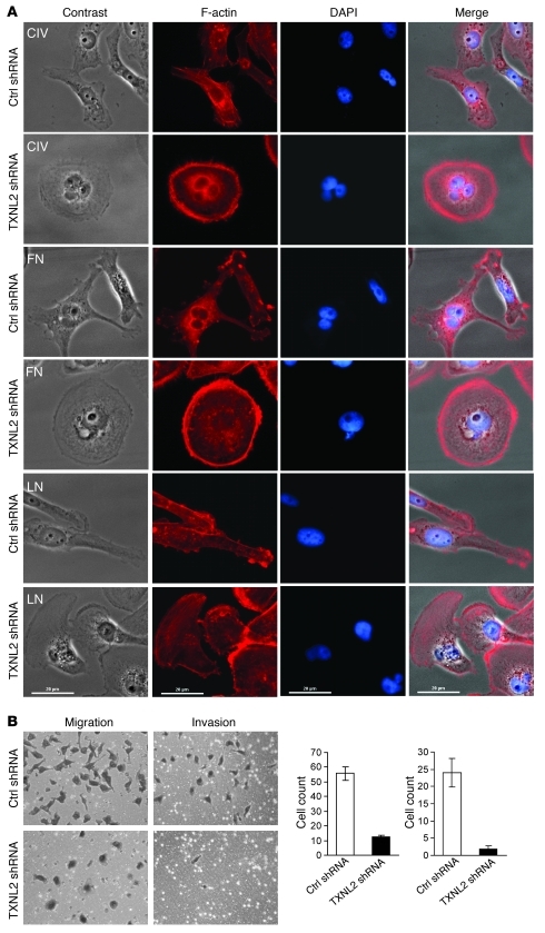
Figure 3. Knockdown of TXNL2 blocks the motility of MDA-MB-231 cells.
(A) Cell morphologies on different extracellular matrix surfaces are shown. Control and TXNL2-KD MDA-MB-231 cells were plated on collagen IV (CIV), fibronectin (FN), and lamin (LN). F-actin was stained with Alexa Fluor–labeled phalloidin. DAPI was used to visualize cell nuclei (original magnification, ×400). (B) Migration and invasion of control and TXNL2-KD cells were measured using transwell chamber assays. Data represent the average cell number from 5 viewing fields (original magnification, ×200).

