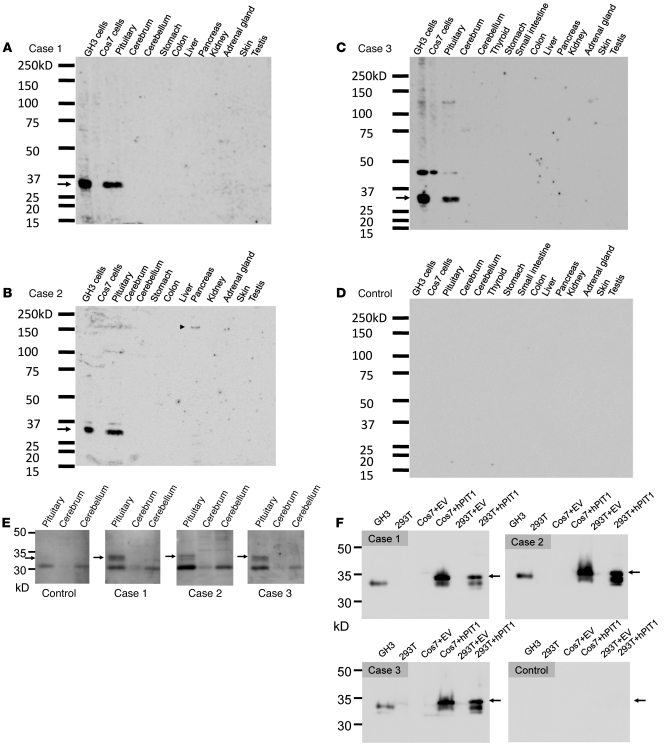
Figure 1. Immunoblotting analysis of mouse tissues and cell lysates.
The patients’ serum was used as a primary antibody (1:1000) to detect the autoantibody. (A–C) The patients’ sera specifically recognized a 33-kDa protein in the lysates from the pituitary and GH3 cells (arrow). (B) The serum of patient 2 recognizes a 150-kDa protein in the pancreas (arrowhead), but the size of this protein does not correspond to that of insulin (58 kDa) or GAD (65 kDa). (C) The serum of patient 3 recognized approximately 45 kDa protein as a nonspecific band in addition to the 33-kDa protein PIT-1. (D) Representative results of the healthy control subjects are shown. Neither the sera of 10 healthy control subjects nor those of 8 patients with pituitary adenoma and 6 patients with hypophysitis recognized the 33-kDa protein. (E) The patients’ sera detected the 33-kDa protein in the lysate from human pituitary. (F) The patients’ sera detected the PIT-1 protein in the lysates from GH3, hPIT-1–expressing Cos7, and 293T cells (arrows).

