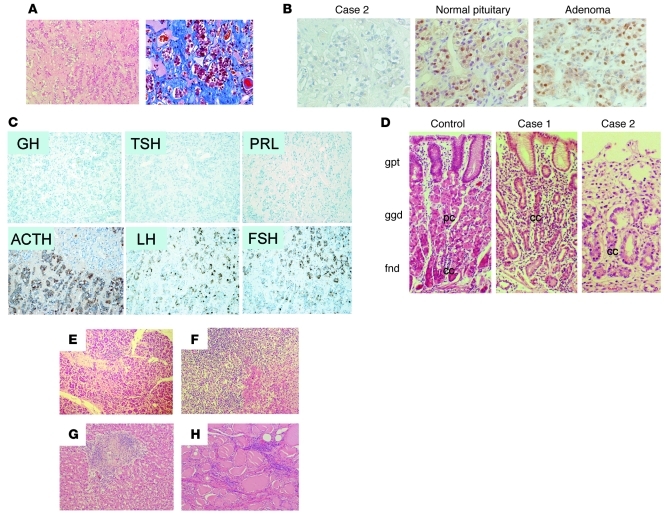
Figure 4. Histological analysis revealed the defect in PIT-1–positive cells and multiple endocrine organopathy.
(A) Microscopic analysis of H&E-stained sections of the pituitary tissue from patient 2. The number of adenohypophyseal cells is markedly reduced. The reduction is accompanied by infiltration of lymphocytes and plasma cells (left panel) and mild fibrosis in the stromal tissue (right panel: Azan staining). (B) Immunohistochemical analysis using an anti–PIT-1 antibody. In contrast to the normal pituitary or pituitary adenoma, no PIT-1–positive cells are observed in the pituitary in the case of patient 2. (C) Immunohistochemistry of the pituitary in the case of patient 2. GH-, PRL-, and TSH-positive cells are absent, despite the presence of ACTH-, LH-, and FSH-positive cells. (D) Histological analysis of the gastric mucosa from control and patients 1 and 2. In contrast to the control, no parietal cells were detected in patients 1 and 2. gpt, gastric pit; ggd, gastric glands; fnd, fundic glands; pc, parietal cells; cc, chief cells. Histological analysis of tissue of the pancreas (E), adrenal gland (F), liver (G), and thyroid (H) from patient 2 revealed inflammatory infiltration and disrupted tissue structure. Original magnification, ×100 (A, C); ×400 (B); ×200 (D); ×100 (E–H).

