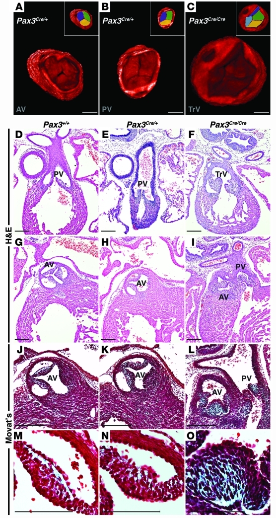
Figure 1. Loss of Pax3 results in abnormal semilunar valve leaflets.
3D reconstruction of OPT images of E16.5 Pax3Cre/+ control embryos demonstrates a trileaflet aortic (A) and pulmonic (B) valve, each with 3 commissures, while Pax3Cre/Cre mutant embryos demonstrate abnormal semilunar valve leaflets. The mutant depicted in C displays a truncal valve with 4 leaflets. Insets in A–C represent identical images with each leaflet pseudocolored. Cross-sectional H&E images through the pulmonic valve leaflets of E16.5 Pax3+/+ and Pax3Cre/+ embryos (D and E) and a Pax3Cre/Cre littermate (F) show thickened leaflets in the mutant. Cross-sectional H&E images through the aortic valve leaflets of E16.5 Pax3+/+ and Pax3Cre/+ embryos (G and H) and a Pax3Cre/Cre littermate (I) show thickened, unequally sized leaflets in the mutant. Examples of a truncal valve (F) and double-outlet right ventricle (I) are shown. Modified Movat’s Pentachrome staining reveals extracellular matrix deposition (blue) in E16.5 control semilunar valve leaflets (J and K) and Pax3Cre/Cre embryos (L), which show an increase in extracellular matrix compared with control leaflets. Higher magnification images of J–L are shown as M–O. AV, aortic valve; PV, pulmonic valve; TrV, truncal valve. Brightness and contrast of OPT images were adjusted using OsiriX software. Scale bars: 100 μm.

