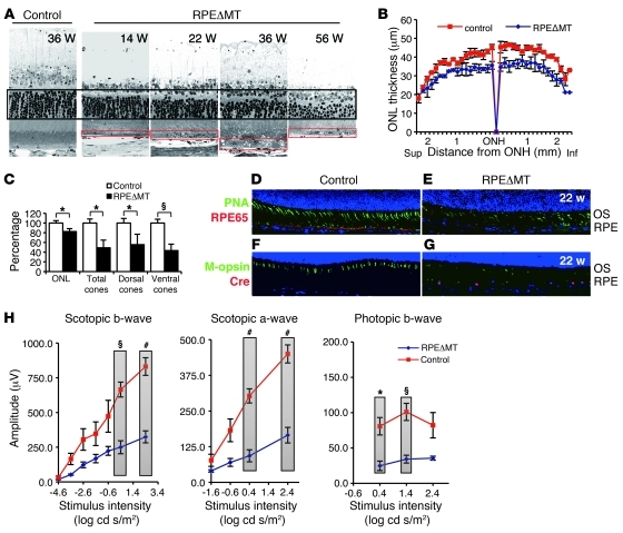
Figure 4. Photoreceptor degeneration correlates with RPE dedifferentiation in albino RPEΔMT mice.
(A) Light micrographs of the posterior retina show a progressive regional loss of photoreceptors reflected by outer nuclear layer (ONL) thickness (black box) and correlating with RPE dedifferentiation (red boxes). Original magnification, ×630. (B) Global assessment of outer nuclear layer thickness in 36-week-old RPEΔMT mice (n = 3) and controls (n = 3). Sup., superior retina; ONH, optic nerve head; Inf., inferior retina. (C) Quantification of mean outer nuclear layer thickness and cones shows a significant loss of rod and cone photoreceptors in 36-week-old RPEΔMT mice (n = 3) compared with that in controls (n = 3, mean value of controls is defined as 100%). (D–G) Immunostaining images from the posterior ventral retinas of 22-week-old mice. Original magnification, ×200. (D and E) Costaining for lectin-PNA (green) and RPE65 (red) demonstrates (E) a striking loss of cones in RPEΔMT mice, which correlates with diminished RPE65 protein. (F and G) Immunostaining for M-opsin (green) and cre (red) shows abnormalities of red/green opsin-expressing cones, which correlate with cre-expresssing cells (G). (H) Electroretinograph demonstrates significantly reduced rod responses (scotopic a-wave and b-wave) and cone responses (photopic b-wave) in 36-week-old RPEΔMT mice (n = 3), compared with those in controls (n = 5). Verticle bars clarify the 2 groups of values used for statistical comparison. Data in H represent mean ± SEM. *P < 0.05; §P < 0.01; #P < 0.001.

