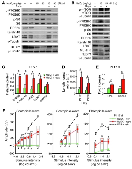
Figure 8. Rapamycin inhibits mTOR/S6K-mediated RPE dedifferentiation and subsequent retinal degeneration induced by NaIO3 in B6 mice.
(A) Immunoblot of proteins from eyecups shows elevated phosphorylation of P70SK6Thr389 and S6Ser235/236 and loss of RPE markers 2 days PI of NaIO3 (2 middle lanes). (B) At day 5 PI, an immunoblot detects persistent activation of P70SK6Thr389 and S6Ser235/236 and diminished RPE markers in isolated RPE cells (middle lane). The effects are inhibited by rapamycin treatment (A, 2 right lanes; B, right lane). (C) Quantification of RPE proteins 5 days PI of NaIO3 (triplicates) shows preservation of several RPE markers after rapamycin treatment (red bars) (normalized to PBS and vehicle-treated controls). (D and E) Quantification of the length of normal retina (RPE and neural retina) at PI days 17 and 20 (D), and outer nuclear layer thickness and cone density at PI day 17 (E). Rapamycin treatment (red bars, n = 6) results in a striking preservation of retinal morphology relative to that of vehicle treatment (green bars, n = 6) in NaIO3-injected mice. In E, the mean value of outer nuclear layer and cones of vehicle-treated mice was defined as 1. Data represent the relative fold increase of the corresponding value obtained from rapamycin-treated, NaIO3-injected mice. (F) Electroretinography demonstrates significantly increased scotopic and photopic responses at PI day 17 in NaIO3-injected B6 mice treated with rapamycin (red, n = 5), compared with those in vehicle-treated NaIO3-injected controls (green, n = 6). The dashed curve represents control mice (PBS + vehicle, n = 7). Verticle bars clarify the 2 groups of values used for statistical comparison. Data represent mean ± SEM. *P < 0.05; §P < 0.01; #P < 0.001.

