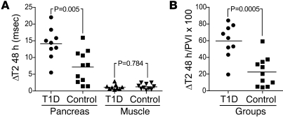
Figure 3. MRI-MNP may be used for the noninvasive quantification of pancreatic changes associated with the development of diabetes.
(A) ΔT2 was measured inside matching ROIs before and 48 hours after infusion of MNPs, reflecting local accumulation of MNPs. Comparing recent-onset T1D patients (n = 9) and controls (n = 11), there was a significant difference in ΔT2 within the pancreas (T1D 14.1 ± 4.7 msec, controls 7.1 ± 4.9 msec) but not within paraspinous muscle (T1D 1.28 ± 0.78 msec, controls 1.23 ± 0.90 msec). (B) A composite index was computed using the formula 100 × (ΔT2pancreas/PVI); (T1D 59.7 ± 20.1, controls 22.5 ± 17.5).

