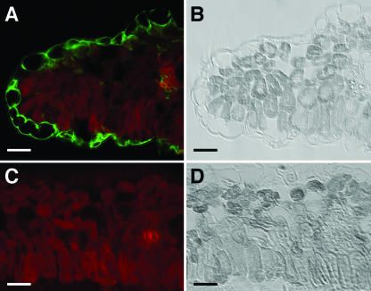
Figure 4.
Immunolocalization of WAKL6. A and C, Sections through 14-d-old leaves were probed with WAKL6 antiserum (A) or preimmune serum (C). Antibody binding was detected with a fluorescein-conjugated secondary antibody. Green fluorescence indicating WAKL6 protein and autofluorescence of chloroplasts (red) in the mesophyll cells are superimposed. B and D, Bright-field images of the sections in A and C, respectively. Bars = 25 μm.

