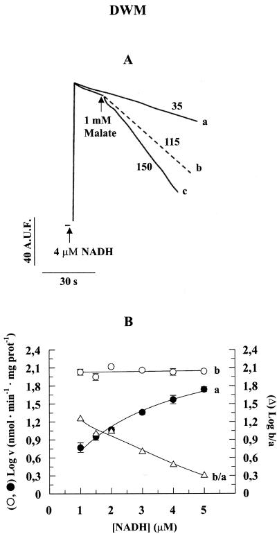
Figure 3.
Fluorimetric measurement of NADH oxidation rate at low NADH concentrations by the external dehydrogenase and by MAL/OAA shuttle in DWM. A, Mitochondria (0.05 mg of protein) were incubated in 2 mL of the standard medium containing 10 EU PH-MDH; then, 4 μm NADH was added, and the fluorescence (λex 340 nm; λem 456 nm) was continuously monitored (trace a). In the case of trace c, MAL was added at the time indicated by the arrow. Trace b was calculated as: c - a. The numbers alongside the traces refer to the nanomoles of NADH oxidized per minute per milligram of protein. A.U.F., Arbitrary units of fluorescence. In this experiment, PH-MDH activity was used giving a ratio of external-MDH activity to MAL-OAA antiporter activity roughly similar to the one due to cytosolic MDH (cMDH) under physiological condition (see Scheme 1). B, The same experiment was repeated using 1 to 5 μm NADH, and the logarithm of the rates (v) of NADH oxidation was reported as a function of NADH concentrations: a, NADH oxidation rate due to NADH DHExt; and b, NADH oxidation rate due to MAL/OAA exchange. The data are reported as mean of three experiments ± se.

