Figure 2.
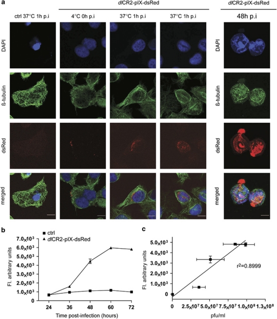
Use of dlCR2-pIX-dsRed to monitor infection, virus replication and exit. (a) Visualization of viral entry and exit. IGROV1 cells were infected with dlCR2-pIX-dsRed on ice for 1 h (0 h p.i. timepoint). Cells were warmed to 37 °C for 1 h, stained with β-tubulin, DAPI and subjected to confocal analysis (left). Scale bar=10 μm. A2780CP cells were infected with dlCR2-pIX-dsRed (MOI 1) and analyzed as above 48 h p.i. (right). (b) A2780CP cells were infected with dlCR2-pIX-dsRed (MOI 1). The emitted fluorescence was measured with a plate reader in 12 h intervals up to 72 h p.i. (c) Scatter-plot analysis of data in B combined with intracellular viral replication as p.f.u./ml showing a correlation between fluorescence and intracellular viral replication (r2 value=0.8999).

