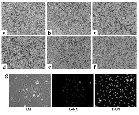
Figure 9.
Loss of spindle cell morphology correlates with loss of KSHV episomes in KSHV-infected HUVEC cultures. At each passage, aliquots of the continuously growing KSHV-infected HUVEC cultures shown in Figure 6a were allowed to reach confluence and maintained for an additional 4 days to allow for complete conversion of infected cells to spindle cell morphology. Shown are cultures seeded at passage 1 (day 2 after infection (a), passage 2 (day 5 after infection) (b), passage 3 (day 7 after infection) (c), passage 4 (day 9 after infection) (d), and passage 5 (day 13 after infection) (e). An image of a mock-infected culture propagated in parallel is shown (f). (g) Cluster of spindle-shaped cells from a culture seeded at day 7 after infection analyzed by light microscopy (LM, left) and immunofluorescence staining for LANA (center). Nuclei were stained with DAPI (right).

