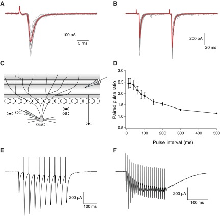
Fig. 1.
Properties of parallel fibers to Golgi cell excitatory postsynaptic currents (EPSCs). A: representative (gray) and averaged traces (red) from a series of 20 consecutive EPSCs evoked by extracellular stimulation of parallel fibers (PFs) in coronal slices. B: in all experiments, EPSCs were evoked with a paired-pulse protocol (10–100 μA, 100–200 μs) with a 50 ms interpulse interval. Representative (gray) and averaged (red) traces from 20 consecutive stimulations show paired-pulse facilitation. C: schematic diagram of representative positions of the stimulating and recording electrode in the coronal slice plane. D: on the average, paired pulse ratios (PPRs) were strongly dependent on the interpulse intervals (n = 9; 20 PPRs per data-point, with error bars indicating SE). E and F: response to tetanic stimulation, averaged over 20 responses, for stimulation at 50 (E) and 100 Hz (F); the latter was used to induce long-term depression of EPSCs.

