
Fig. 1.
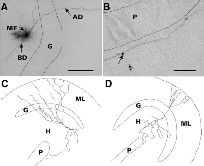
Morphology of representative hilar ectopic granule cells (HEGCs). Cells were filled with biocytin during whole cell patch-clamp recording, fixed slices were cut into serial 60-μm-thick sections, biocytin was visualized with nickel-intensified avidin-horseradish peroxidase-diaminobenzidine (Vectastain Elite ABC kit, Vector Laboratories, Burlingame, CA), and cell morphology was reconstructed with use of Neurolucida (MicroBrightField, Williston, VT). Criteria for confirmation of HEGC identity included hilar location of the soma, apical dendrite(s) penetrating into or directed toward the dentate molecular layer, and axon with giant boutons in area CA3 and extensive branches within the hilus. A: the apical dendrite (AD) of this HEGC crosses the granule cell body layer (G) and penetrates the molecular layer. The mossy fiber (MF) branches within the hilus. This cell also has a basal dendrite (BD) directed into the hilus. Scale bar, 500 μm. B: HEGC mossy fiber courses through stratum lucidum of area CA3 adjacent to the pyramidal cell body layer (P). Arrow indicates a giant bouton from which a filopodium originates. Scale bar, 200 μm. C: reconstructed morphology of an HEGC from which miniature inhibitory postsynaptic currents (mIPSCs) were recorded. D: reconstructed morphology of an HEGC from which miniature excitatory postsynaptic currents (mEPSCs) were recorded. The apical dendrites of both cells reached the outer edge of the dentate molecular layer (ML) and the cell in D had a short basal dendrite directed into the hilus (H). The main branch of the mossy fiber reached stratum lucidum of area CA3.