
Fig. 1.
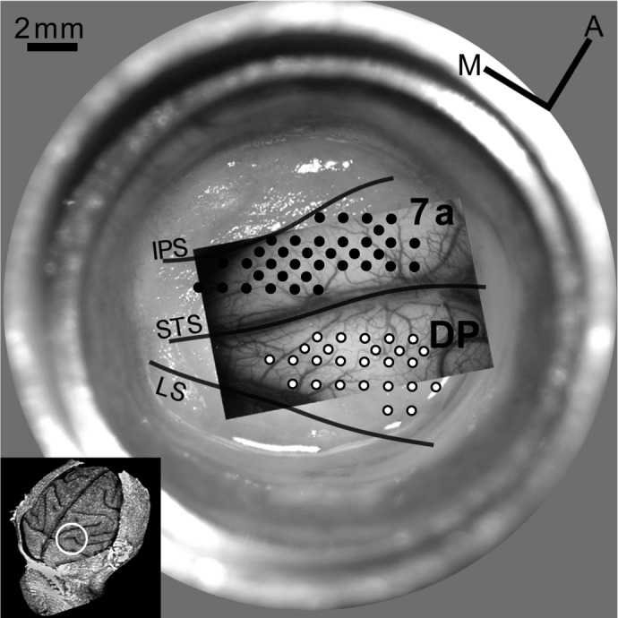
Recording sites in optical chamber (M1R). Image of blood vessel pattern (green light, 540 nm) and grid with recording sites overlaid on chamber picture with regrown dura. Area 7a is located between intraparietal sulcus (IPS) and superior temporal sulcus (STS; filled circles mark recording sites); the dorsal prelunate (DP) is located between lunate sulcus (LS) and STS (open circles mark recording sites). Grid, 1-mm spacing. Small inset shows anatomical overview of the right hemisphere of M1R reconstructed from structural MRIss. White circle marks location of recording chamber relative to the sulcal pattern.