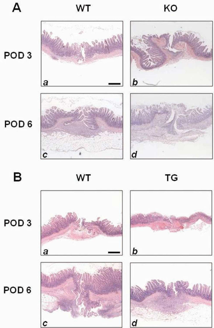
Figure 2.
Histology of anastomoses. Shown are representative H&E stained histological images of ileal anastomoses on POD 3 and 6. A) Histology of HB-EGF KO mice and their WT C57BL/6J × 129 counterparts ; B) Histology of HB-EGF TG mice and their WT FVB counterparts. Magnification 20×. Scale bar = 100µm.

