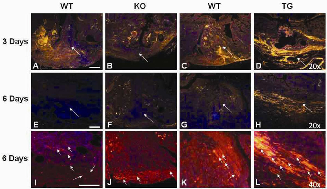
Figure 5.
von Willebrand factor (vWF) immunohistochemistry of anastomotic sites. Shown are representative photomicrographs of vWF immunostaining of the anastomotic sites. Magnification: A-H, 20×; I-L, 40×. Red, vWF (Cy3); blue: DAPI, nuclear staining; WT, wild type; KO, HB-EGF knock out mice; TG, HB-EGF transgenic mice; * p<0.05. Long white arrows indicate the anastomotic sites. The small white arrows in high power panels I-L indicate mature blood vessels containing lumens. Scale bar = 100µm.

