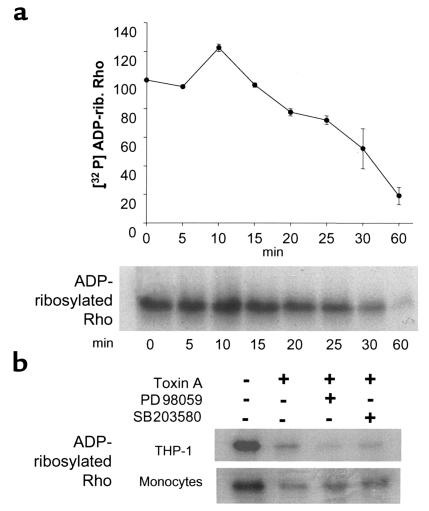
Figure 6.
(a) Rho glucosylation is detected after MAP kinase activation by toxin A. The time course of Rho glucosylation was measured in THP-1 cells exposed to toxin A (100 nM). After the indicated periods of time, cell extracts were incubated with C. botulinum C3 exoenzyme and [32P] NAD to ADP-ribosylate Rho. Proteins were then separated by 12% SDS-PAGE, and ADP-ribosylated Rho was measured by autoradiography and densitometry (n = 3). Rho glucosylation was detected 15–20 minutes after toxin A addition and was complete after 1 hour. Moreover, a 20% increase in ADP-ribose incorporation was observed after 10 minutes of toxin exposure. Rho glucosylation was thus detected after MAP kinase activation that was evident after 1–2 minutes (Figure 1). (b) Rho glucosylation is independent of ERK and p38 activation. THP-1 cells and monocytes were pretreated with 20 μM PD98059 or 10 μM SB203580 for 30 minutes and then exposed to toxin A for 1 hour. The ADP-ribosylation assay was performed as described above. Blocking the ERK and p38 pathways did not prevent Rho glucosylation by toxin A.

