Fig. 2.
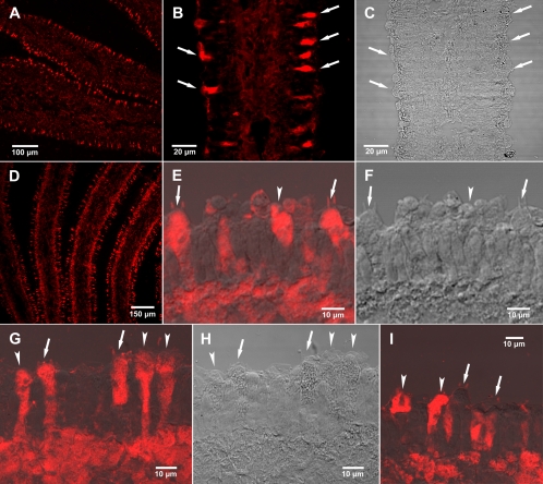
Amanitin immunostaining of lamellae (gills) of A. bisporigera observed by confocal laser scanning microscopy (CLSM). (A) Low-magnification CLSM view showing cross-section of two lamellae and part of a third. (B) Higher magnification of a single lamella by CLSM. (C) Same gill section as in panel B viewed by differential interference contrast (DIC). (D) A different basidiocarp, showing a cross-section of three lamellae and parts of two others by CLSM. (E) Antiamanitin immunostaining of basidia (arrows), indicated by the sterigmata (the two slender projections), sterile cells (arrowheads), and the subhymenium (CLSM superimposed on DIC). Sterigmata are the slender projections from the basidia that bear the basidiospores, which are no longer present in these sections. Sterile cells are the cells in the hymenium that, in Amanita, resemble basidia but do not bear spores. (F) Same section as in panel E viewed with DIC alone. (G) Antiamanitin immunostaining of hymenium and subhymenium showing basidia (arrows) with sterigmata and sterile cells containing amanitin (CLSM superimposed on DIC). (H) Same section as in panel G observed by DIC. (I) Basidia (arrows) showing absence of amanitin and adjacent sterile cells (arrowheads) showing presence of amanitin (CLSM superimposed on DIC).

