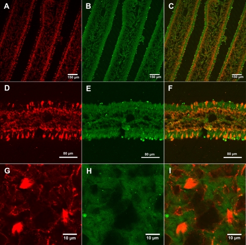
Fig. 4.
Dual immunostaining of A. bisporigera lamellae using antiamanitin and antiactin antibodies. (A to C) Lamella cross-sections, showing amanitin staining, actin staining, and the merge of the two, respectively. (D to F) Higher-magnification views of a cross-section of a lamella (same staining order as for panels A to C). (G to I) Higher-magnification views of a cross-section of a mature pileus.

