Fig. 1.
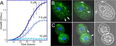
Wild-type T. thermophila cells are dinitroaniline sensitive. (A) Growth of wild-type T. thermophila at 30°C in medium with 0, 7.5, or 15 μM oryzalin. Wild-type T. thermophila cells showed reduced growth at 7.5 μM and complete inhibition at 15 μM. (B and C) Microscopy of T. thermophila cells grown in the absence (B) or presence (C) of 7.5 μM oryzalin indicated that many microtubule structures are disrupted by dinitroaniline treatment. T. thermophila cells were labeled with antitubulin antibody (green) and 4′,6-diamidino-2-phenylindole (DAPI, which labels the nuclei blue). The left panels show merged blue and green fluorescence, the middle panels show the fluorescence image merged with a phase-contrast image, and the right panels show a phase-contrast image alone. (B, left) Normal T. thermophila cell covered with tubulin-containing cilia, but also with underlying microtubules, such as the cortical microtubules (arrows). (B, middle and right) T. thermophila cilia are visible by both tubulin immunofluorescence and phase imaging (arrows). (C, middle) Oryzalin-treated T. thermophila cells lose most or all cilia, making the underlying cortical microtubules (arrows) more visible.

