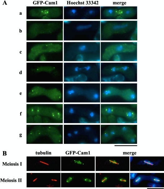
Fig. 1.
Cellular localization of Cam1 during mating, meiosis, and sporulation. The homothallic haploid strain AI248 harboring a gfp-cam1 fusion gene was cultured on SSA medium to induce the sexual cycle. (A) Cells were stained with Hoechst 33342 to visualize nuclei. Developmental stages shown are as follows: row a, before karyogamy; row b, horsetail stage; row c, before meiosis I; row d, meiosis I; row e, interphase; row f, early meiosis II; row g, after meiosis II. Bar, 10 μm. (B) Cells were fixed during meiosis. Microtubules and nuclei were visualized by anti α-tubulin antibody, TAT1 (red) (32), and DAPI (blue), respectively. Bar, 10 μm.

