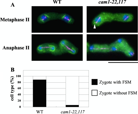
Fig. 3.
Initiation of FSM assembly in the cam1-22,117 mutant. (A) The nascent FSM was visualized by GFP-psy1 (green). A cam1+ strain (YM20) and a cam1-22,117 strain (AI52) were cultured on MEA sporulation medium and doubly stained with anti-α-tubulin antibody, TAT1 (magenta), and DAPI (blue). The arrowhead indicates a very short FSM-like structure. Bar, 10 μm. (B) Quantitative assay for zygote formation with an FSM. Only zygotes that assembled meiosis II spindles were counted.

