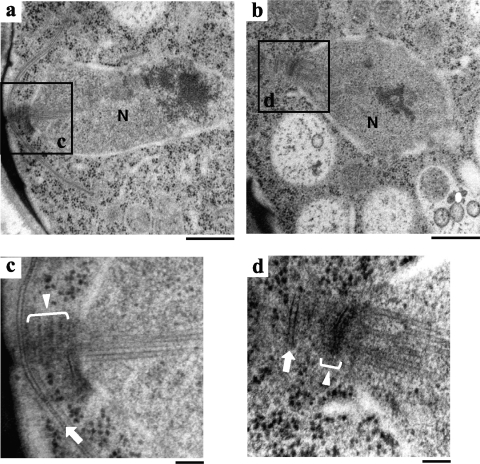
Fig. 4.
Fine structures of wild-type and cam1-22,117 strains during meiosis II. Cells were fixed after incubation on MEA sporulation medium for 1 day. Electron microscopic images of the wild-type (a and c) and cam1-22,117 (b and d) strains. Panels c and d are magnified images of the boxed regions in panels a and b, respectively. N, nucleus; white lines with triangles, the SPB; white arrows, the expanding FSM (c) and the abortive FSM (d). Bars, 500 nm (a and b) and 100 nm (c and d).

