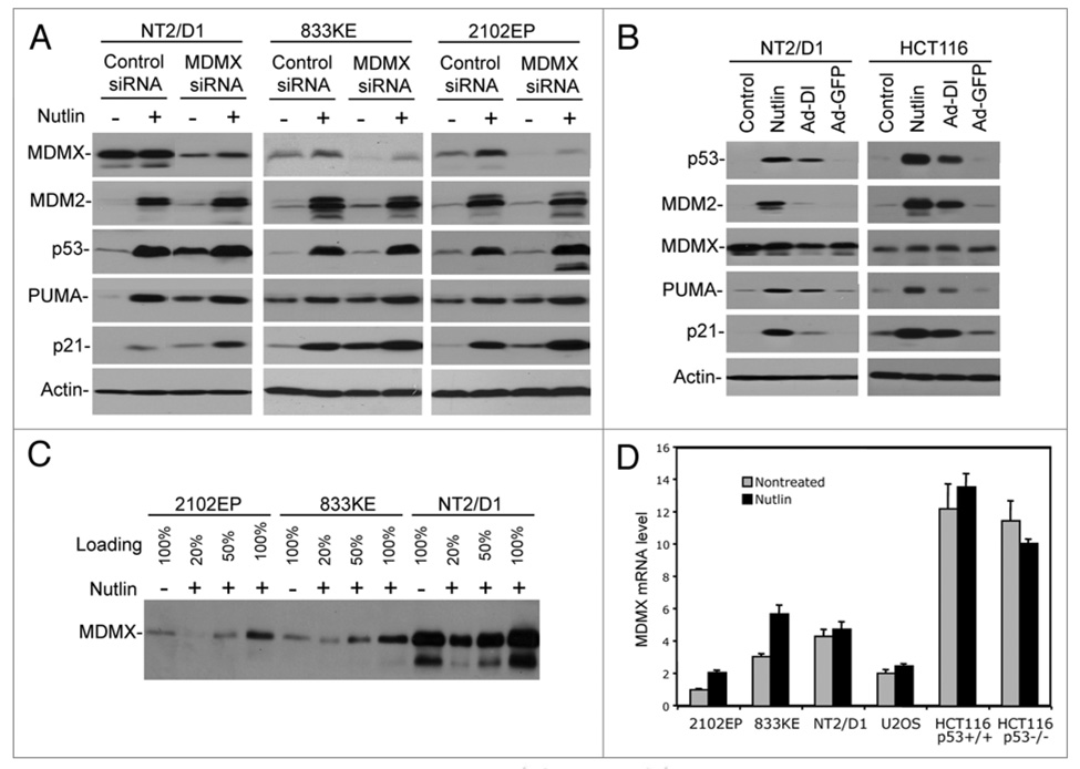
Figure 2.
MDMX contributes to p53 inactivation in TGCT cells. (A) Cells were transfected with MDMX siRNA or control siRNA for 48 hrs and then transfected again with the same amount of siRNA, and 19 hrs later cells were treated with 5 µM Nutlin for 8 hrs. Expression levels of indicated markers were analyzed by western blot. (B) Cells were infected with adenovirus Ad-DI or Ad-GFP (MOI = 300) and collected 48 hr post-infection. Expression levels of indicated markers were analyzed by western blot. (C) Cells treated with 5 µM Nutlin for 18 hrs were analyzed by western blot at different loading levels. (D) Cells treated with 5 µM Nutlin for 18 hrs were analyzed by quantitative RT-PCR to determine the level of MDMX mRNA.

